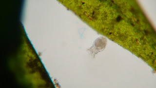
The final week rolled around and I missed it. So I had to catch the emergency week just in case you missed anything. I ended up having a doozy of a last day as my aquarium seemed to have to life. The Euchlania still had the highest amount of creatures at 17, but the Cyclops seemed to be playing catch-up as I counted their number at 6. The diving beetle was of course still around exerting his reign over the tiny aquarium and the green beetle was still nowhere to be found. As I began looking for new creatures I immediately found one in the algae covered Amblestegium Varium moss. It was a insect larvae of a midge, Chrinomus sp (Pennak). It was moving all over the place eating as much of anything as it could. It was really interesting being able to view the larvae literally swallow things whole and watch them go down it's digestive track. The larvae was so long and so fast that I was unable to successfully capture it in one take.
Chrinomus sp. Austin Troutt
Chrinomus sp. Austin Troutt
Also in these pictures you can see just how much the algae has taken over the small environment. Doing a bit of research I was able to find out that the algae is the Golden Algae or Chrysophytes (Forest).
That basically wraps things up for this project. I gotta say this was a really fun project and for a miniscule moment I felt like a real scientist. I hope there are projects like this in Botany II.
References:
Forest, HS. Handbook of Algae. The University of Tennessee Press. 1954. page 278
Pennack, RW. Fresh-water Invertabrates of the United States. John Wilson and sons. 1989 page 687
Microaquarium
Wednesday, November 27, 2013
My fourth week of observation proved to be very exciting as I was able to identify a lot of creatures. The Euchlania have more than doubled as it seemed that I saw one with every turn of the microscope.
Euchlanis sp. Austin Troutt
The diving beetle seems to still be doing fine, however, there is no sign of the green beetle. At this point I'd like to take a moment of silence for the green beetle as his fate is all but certain. I wish I could have identified him before he was decomposed but some things are better left a mystery. However, with death comes life I was able to identify a new species in my aquarium. Swimming around near the bottom I was able to identify a single water flea, Cyclops bicuspidatas (Patterson). It was quite fast and seemed a bit photo shy, but I was able to take a few pictures of the beast before it would dart away.
Cyclops sp. Austin Troutt
Cyclops sp. Austin Troutt
The algae seems to have stopped growing as the layer it provides still is about as thick as it once was.
References:
Patteson, DJ. Free-living Fresh-water Protozoa: A color guide. Manson Publishing. Boca Roton, FL. 1992. Page 163
In my third week of observations, I observed a
lot more diversified life in my aquarium. A food pellet had been dropped in the
aquarium in order to attract the creatures and I was able to successfully identify
a Euchlanis sp. Rotifers. Euchlania are filter feeders, meaning they filter particles
out of the water (Patterson). I was able to identify 4 Euchlania. The diving
beetle is still alive and doing well and so is the small green beetle though I
still cannot slow it down to identify it. I was not able to find any planarian
this time around. The algae have grown exponentially and now the aquatic plants
have a thick layer of algae on them.
Euchlanis sp. Austin Troutt
References:
Patterson, DJ. Free-living Fresh-water Protozoa: A color guide. Manson Publishing. Boca Rotan, FL. 1992 page 133
In the second week, I observed a lot more creatures in my
aquarium. I was able to successfully observe a planarian due to its shape and its
skeletal structure, but was unable to capture on camera. The planiria was able
to move by beating its cilia on the ventral dermis which allows the flatworm to
swim in the water (Panteleimon). So far that is the only planarian I have seen.
Researching, I was able to identify the aquatic beetle that I saw in the first
week. I was able to identify that the beetle was a diving beetle called
Scarodytes Halensis, due to the beetle having the black dots on its back resembling
diving beetles and the general size matched (Pennack). I am still unable to
identify the small green beetle as I cannot make a proper identification with
my eyes and it moves too fast in the water for me to take a concentrated
picture of it. I also observed that the aquatic plants Utricularia gibba and
Amblestegium Varium had a lot of algae of some sort growing on them. It
seemed to be a small layer that had just started, but it was not there last
week.
References
Pennack, RW. 3rd edition Fresh-water Invertabrates of the United States. john Wiley and sons. 1989. pg 456
Panteleimon Rompolas, Ramila S. Patel-King, Stephen M. King
Chapter 4-Schmidtea Mediterranea: A Model System for Analysis of Motile Cilia
Methods in Cell Biology, Volume 93, 2009, page 82
Tuesday, November 26, 2013
For my Micro-aquarium project i used the water and soil from Meads Quarry, Island Home Ave to fill my container 3/4 full by sucking it up with a pipette (McFarland 2013). I than added the aquatic plants ,Utricularia gibba from Spain Lake on Camp Bella Air Road and Amblestegium Varium from a spring in Carters Mill Park on Carter Mill road ,to my container and it rose to about 4/5 full (McFarland 2013). When adding the soil from Meads Quarry I accidentally picked up an aquatic beetle and added it to my aquarium. It is the largest of any of the creatures so far as you don't need a microscope to see it. I also picked up a tiny green beetle as well that only an expert could identify with the naked eye, but too large for a microscope. While looking through my aquarium i wasn't able to see much of any other creatures, however, i was able to clearly see the plant cell structures in both of my aquatic plants.
References:
McFarland, Kenneth [Internet] Botany 111 Fall 2013. [November 27,2013]. Available from http://botany1112013.blogspot.com/
References:
McFarland, Kenneth [Internet] Botany 111 Fall 2013. [November 27,2013]. Available from http://botany1112013.blogspot.com/
Subscribe to:
Posts (Atom)

.JPG)


